Page Links:
A. The Retina:

The retina consists of:
the fovea: located in the centre. This is a very small area (< 1 mm2) that contains three types of cone photoreceptors (for red, green and blue). This small area provides for sharp and colour vision. (memory trick; cone = colour)
the peripheral retina which contains only rod photoreceptors. These rods only sense black and white but are more sensitive than the cones.
There is also a blind spot, located in the inferior and nasal quadrant of the eye where the optical nerves exit the eye on their way to the brain.

Photoreceptors:
Both types of photoreceptors (rods and cones) share the same plan:
- At one end is a stack of “shelves” which are really infoldings of the plasma membrane. These shelves contain millions of photo pigment molecules (such as rhodopsin).
- The second part (or segment) contains the molecular machinery for the cell (mitochondria etc).
- The third part contains the nucleus
- At the other end is the synapse that connects the receptor cell to other nervous cells in the retina.
The difference between the two cells is that in the rods, the shelves are of the same size whereas in the cones, the shelves diminish in size further away from the cell body, hence its shape and its name.

1. Rhodopsin:
this is the molecule that is waiting on the shelves to be excited by a passing photon. In a normal situation, there will be millions of rhodopsin waiting.
2. Excitation:
Once excited by a light ray, the collision with the photon will cause one chemical bond in rhodopsin to change from a -trans to a -cis configuration. This is very fast. This molecule is now called bathorhodopsin (the names are not really important here).
3. Unstable:
This new molecule is very unstable and changes spontaneously into the next molecule, lumirhodopsin, which is still unstable and changes into the next (metarhodopsin I) which finally stabilizes as metarhodopsin II.
4. Delay:
All these spontaneous transformations also take sequentially more time (from pico- to micro- to milliseconds). This is necessary for the metabolic processes in the cell (which takes milliseconds) to react to this excitation
5. Restoration of Rhodopsin:
The final stable meta-rhodopsin II is converted through scotopsin back to rhodopsin. This takes time (minutes) and energy (ATP).
6. Vitamin A:
The rhodopsin molecules are derived from vitamin A. If there is not enough vitamin A (deficient diet) then the person becomes gradually less sensitive to light (night blindness).
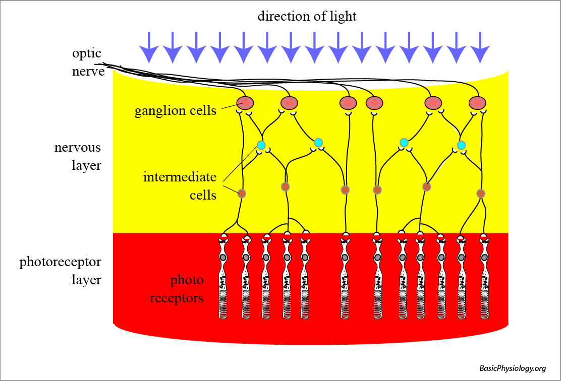
The retina has two layers:
- the photoreceptor layer: which consists of rods (shown in this diagram) or cones.
- the nervous layer: the receptor cells do not connect immediately to the brain cells. Instead, they interconnect with other nerve cells. These intermediate cells already process the signals before communicating with the ganglion cells. The axon of the ganglion cells then combines to form the optic nerve.

1.
The blind spot is located where the axons from the ganglion cells leave the eye ball. Because the nervous layer is on top of the photo-receptor layer, the nerve has to go through this photo receptor layer to reach the sclera and leave the eye. Therefore, at that site, the photoreceptor cell layer is interrupted, hence the ‘”blind” spot.
2.
Note that if the photo receptor layer had been at the other side of the nervous layer, and in front of the light rays, that then, there would have been no blind spot!

The ciliary muscle
also controls the iris and therefore the size of the pupil. It actually consists of two muscles; one outer muscle which is oriented in the radial direction and a second inner muscle that is oriented in the circular direction.
Miosis:
When the circular muscle contracts, the hole (= pupil) within the muscle becomes smaller. This works like a sphincter that you can see in other parts of the body (in the gut or the blood vessels for example).
This is actually a famous reflex (pupillary light reflex) that doctors often use when shining a bright light into the eye to check whether the patient is still alive. This reflex is controlled by the parasympathetic nervous system.
Mydriasis:
is the opposite action (dilatation of the pupil) which is caused by contraction of the radial muscle. This happens in dim light allowing more light into the eye.
This contraction is controlled by the sympathetic nervous system.
Chemical Adaptation:
Dark adaptation:
When you step from a bright room into a dark one, then initially, sensitivity to vision is much reduced. But with time, the eyes become more sensitive and one sees better in the dark.
Light adaptation:
is the opposite of dark adaptation. When you step from a dark to a bright room, then all the rhodopsin molecules will be activated.
This is because, when you stepped from the bright room, a lot of the rhodopsin had been activated and were being used in the cycle. These molecules were therefore not available to pick up the few photons in the dark room.
But with time (up to minutes), the molecules revert back to the rhodopsin shape and the amount of rhodopsin molecules increases. This makes the eye more sensitive.
Dark adaptation:
The pupil dilates (with the radial fibres) to allow more light into the eye (sympathetic reflex).
Light adaptation:
The pupil constricts (with the circular fibres) to reduce the amount of light into the eye (parasympathetic reflex).
Central vision:
This is the vision as captured by the fovea. Most of the photoreceptors in the fovea are cones and practically every cone has its own nerve to the brain. Therefore, the image is sharp and in colour. The whole eye is built to project the image sharp (in focus) onto the fovea.
Peripheral Vision:
The remainder but largest part of the retina is covered by the rods, which are light sensitive but not colour sensitive.
In fact, the rods are more light-sensitive than the cones. For example, at night when you are outside, and you look at a faint star, you may see it better when you don’t look at it straight but at a slight distance from the star.
Warning System:
The rods in the peripheral retina, and the nervous system attached to them, are especially sensitive to movements. Therefore, when something moves, in the “corner” of the eye, it attracts our attention; we turn our eye towards the source of the movement and see it sharp and in colour.
Tunnel Vision:
In some patients, who have their peripheral vision destroyed, the lack of a peripheral vision is striking. They can still see clearly and in colour with their fovea but they are often involved in road accidents, as they have not seen other cars moving into their path from other directions. They lack an early warning system. (Chronic glaucoma for example).