A. Deafness (Hearing Abnormalities):
- Deafness can be caused by an abnormality in the outer ear, the middle ear or the inner hear.
2.
3.
Abnormalities in the cochlea (the inner ear) or in the nerves are called Nerve deafness or Sensorineural deafness.
4. Audiogram:
An audiogram is a measurement of how well we can hear. This is tested by producing sound at different frequencies and at different amplitudes and plotted in a graph (= audiogram). Furthermore, we can test, separately, air and bone conduction.
5.
In air conduction, sound is perceived through the outer ear, the middle ear and the inner ear.
In bone conduction, sound is perceived by creating sound waves through the skull, thereby bypassing the outer and the middle ear and only stimulating the inner ear.
6.
- Audiogram:
An audiogram is a graph that plots the threshold levels for hearing sounds at different frequencies, usually from 125 to 8000 Hz. These levels are expressed in decibels (dB) on a logarithmic scale. Zero dB represents threshold for normal healthy people.
2.
3. Decibel:
is the unit of sound on a logarithmic scale. It is actually the ratio between the actual sound and the threshold for sound which is set at 0 dB. A whisper produces a sound of 20-30 dB, a conversation 40-50 dB, a vacuum cleaner produces 80 dB noise, a truck rumbles at 90 dB, and a civil defense siren blasts at 130 dB which is close to the threshold of pain.
Hz (= Hertz; a former German scientist):
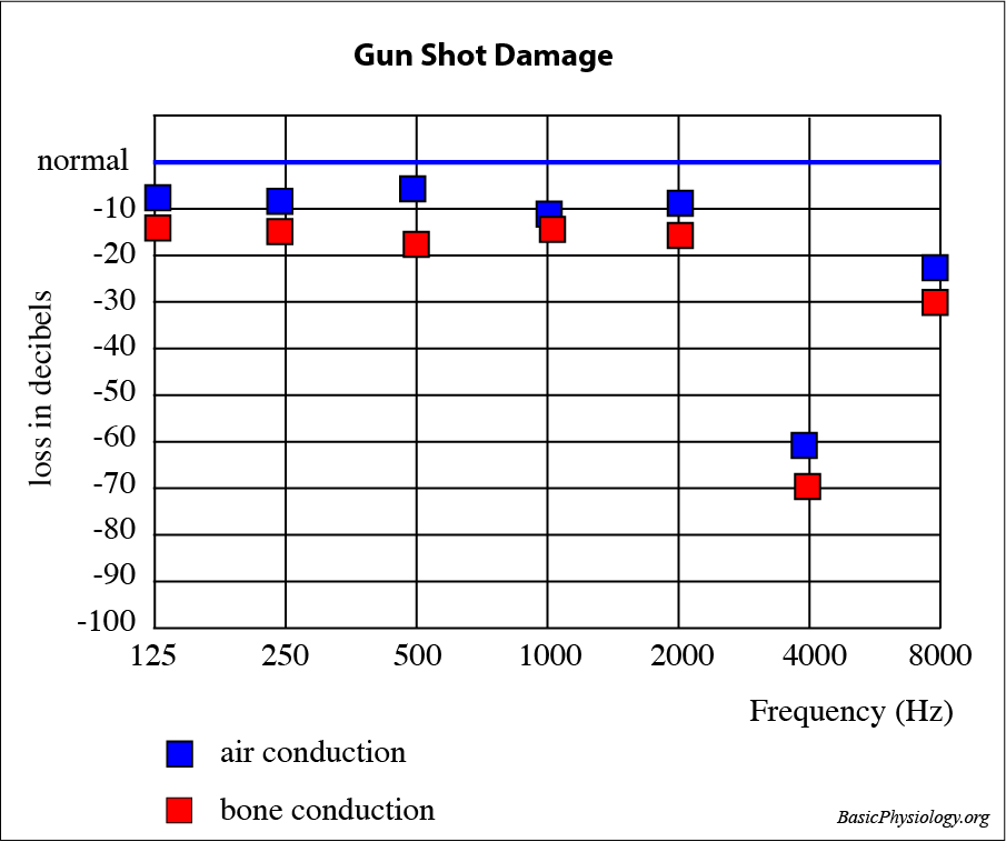
- Gun-shot damage:
In this patient, air and bone conduction is impaired at a specific frequency; in this case at about 4000 Hz. Since bone and air conduction are both equally affected, it is a sensorineural deafness. A damage at one frequency means that a small portion of the basilar membrane is damaged; usually by being exposed repeatedly to a high-volume noise of a specific frequency; for example, the cocking of the head and the ear close to the barrel of a rifle (hence the name!). Nowadays, such damages often occur when people work in environments where there is a lot of (industrial) noise such as airports but also disco’s etc.
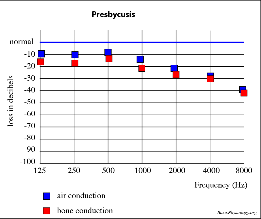
2. Presbycusis:
In this patient, the hearing for higher frequencies is impaired, whereas it is ok at the middle and lower frequencies. Both the air and the bone conduction have declined at the high frequencies. This implies that the problem is located in the inner ear; the cochlea or the nerve. Usually, this is due to the wear and tear in the organ of Corti (= old age!).
3. Impacted Wax:
In this case, the external auditory canal is blocked by old and hardened wax. This is a typical conduction problem. The bone conduction is therefore normal. A ruptured tympanic membrane would produce a similar audiogram.
4. Otosclerosis:
In this patient, bone has accumulated in the middle ear thereby impeding the movements of the ossicles. This is a conduction problem as there is no problem with the cochlea. So, the measured bone conduction is normal but his conduction is impaired. The same type of pattern could be obtained in the case of an otitis media (= middle ear inflammation).


