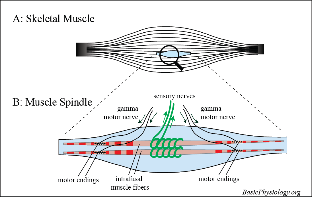
Muscle spindles are stretch receptors that are located/distributed in the belly of skeletal muscles.
2.
They primarily detect changes in the length of the muscles which is (of course) very useful to know, for the brain!
For this function, special muscle fibres are located inside the spindle. These are called intrafusal fibres (and the ‘normal fibres outside the muscle spindles are now called extrafusal fibres).
3.
4.
Around these intrafusal muscle fibres, there are loops of sensory nerve endings wrapped around the fibres. These detect the length/tension in these intrafusal fibres and send that information to the brain.
5.
In addition, the muscle fibres in the muscle spindles can also be stimulated, just like the extrafusal fibres, with efferent nerves, called gamma motor neurons.
6.
As we will see later, these innervations may influence the sensitivity and the function of the muscle spindles.
7.
How many muscle spindles are there in a typical skeletal muscle? Not many, about 25 to 100, on average, in a single muscle.
8.
But there is a large variation in muscles. Some muscles, such as muscles in the legs or the arms have quite a lot of muscle spindles whereas other muscles such as the facial muscles have little or even no muscle spindles at all.
9.
We don’t really know yet what determines this variation. Amount of cerebral control is of course important. But why facial muscles don’t have muscle spindles at all, no idea!
The function of the muscle spindle is to constantly measure the length of the skeletal muscle, which it does with the sensory endings wrapped around the intrafusal fibres.
2.
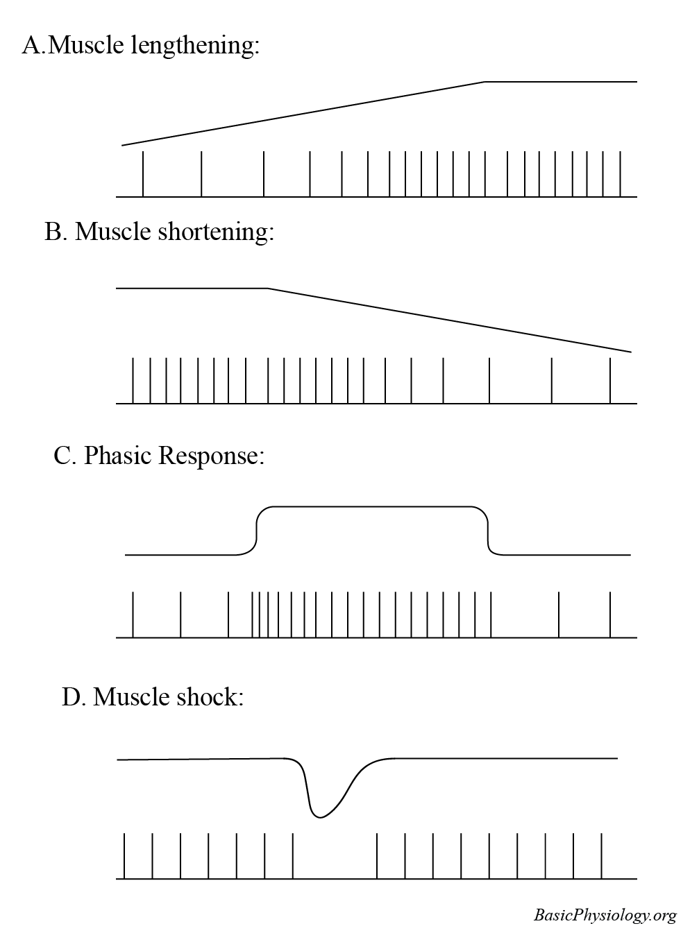
At rest, the muscle spindles discharge a constant rhythm of action potentials that propagate to the brain.
3.
When the muscle is gradually stretched, the action potential frequency gradually increases (panel A) whereas, when the muscle is shortened, the opposite occurs (B).
4.
If there is a sudden increase in the length of the muscle (a jerk) then the firing of the action potentials will suddenly increase and then gradually settle back to that particular length (C).
5.
And, when the muscle is suddenly shortened, the opposite occurs (D).
6.
Btw, why are the gamma motor nerves innervating the intrafusal fibres?
7.
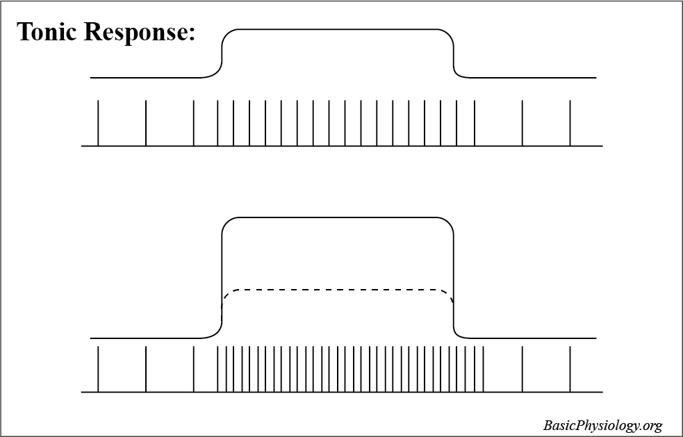
These gamma motor nerves are constantly sending action potentials to the intrafusal fibres which makes these fibres always (slightly) contracted.
8.
That is important because it means that the fibres are constantly alert to changes in the skeletal muscle length.
9.
Suppose, that the intrafusal fibres were not constantly contracted but just hang loose in the spindle, then they would not detect any change in the skeletal muscle. In other words, constant contraction keeps these fibres alert all the time!
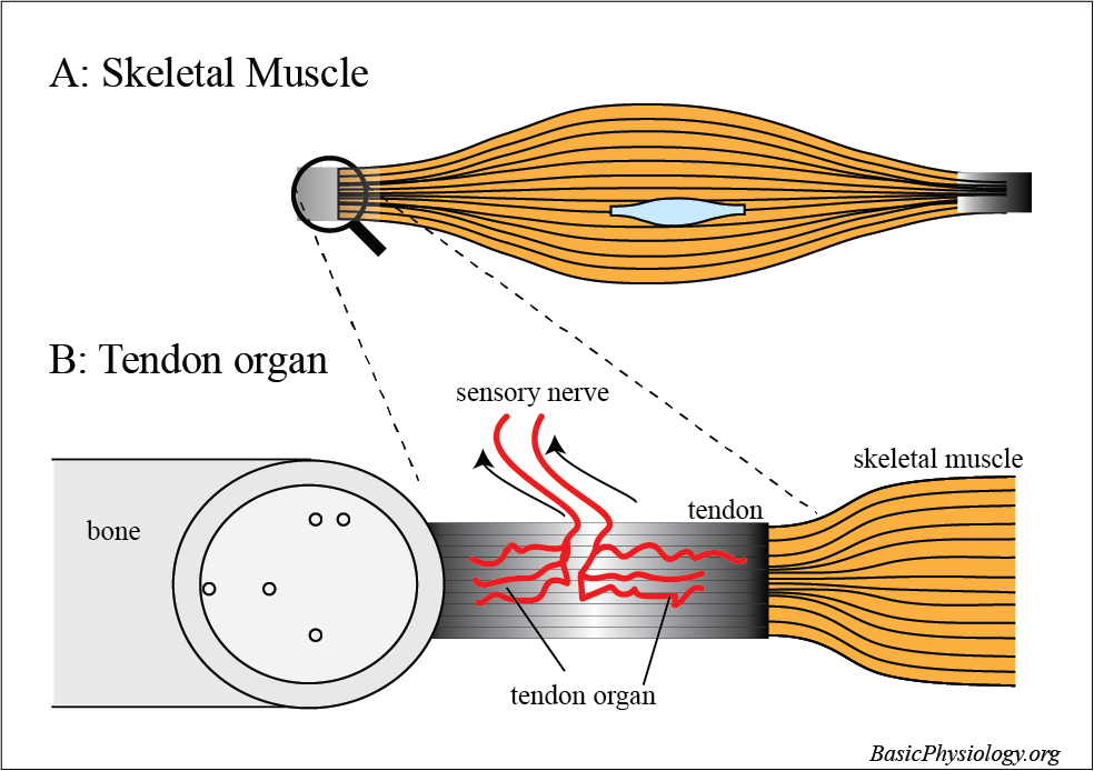
And then, we have another sensor in the skeletal muscles or, rather, in the tendons that fix the muscles to the skeleton.
2.
The tendon organ records the tension in the attached muscle; irrespective whether this is induced by active contraction or otherwise.
3.
The amount of tension is ‘translated’ into the frequency of action potentials that are then send to the brain. The higher the tension, the higher the number of action potentials.
4.
Btw, the tendon organ is sometimes also called the Golgi tendon, since Golgi was the first person to describe this structure (born in 1843!).
5.
Remember Golgi? Yes! He discovered and described the function of the Golgi body; a group of vesicles located in many cells that collect small molecules into vesicles for further transportation (see A.2.1.F.).