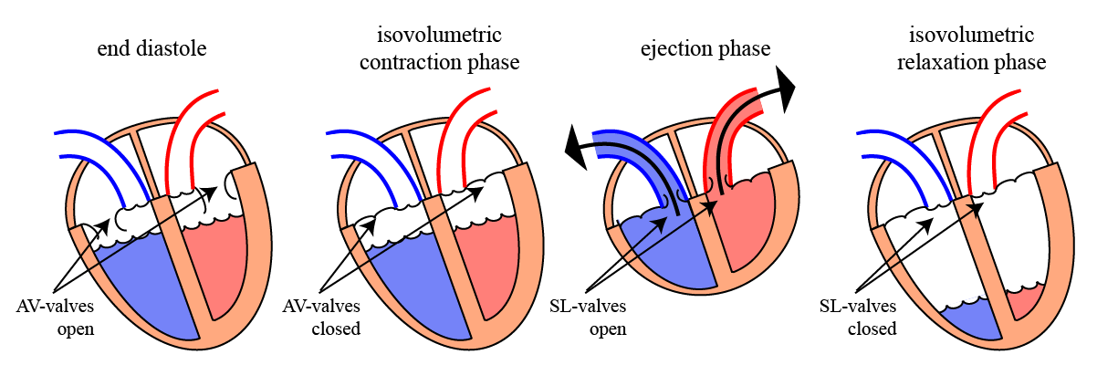
Aim: How does the heart pump the blood into the arteries?

A. Role of the Cardiac Valves:
1.
Remember the structure and location of the cardiac valves?

The cardiac valves play a crucial role in the function of the heart. They make sure that the blood flows in the right direction.

The valves are located between the atria and the ventricles (= the atrio-ventricular valves) and between the ventricles and the arteries (= the semi-lunar valves).
(Semi-lunar = half moon shaped) (RA = right atrium; LA = left atrium; RV = right ventricle; LV = left ventricle).
2.

There are, as we have seen, four different valves:

  1. the tricuspid valves: located between right atrium and right ventricle,
  2. the mitral valves: located between left atrium and left ventricle,
  3. the pulmonary valves: located between right ventricle and the pulmonary artery,
  4. the aorta valves; located between left ventricle and the aorta.
3.

There is no machinery that makes the valves open and close. Instead, it is the blood pressure alone that determines whether the valves are open or closed.

4.

If the blood pressure in the atria is higher than in the ventricles then the AV-valves are open. But, if the pressure in the ventricles is higher than in the atria, then the valves close.

B. Atrial Systole:
1.
During diastole (at rest), the AV-valves are open and the semilunar (SL) valves are closed.
2.
During this phase, the AV-valves are open because blood flows from the atria (higher pressure) into the ventricles (lower pressure). The SL-valves are closed because the pressure in the arteries is (much) higher than in the ventricles.
3.
As we have seen before, the sinus node will initiate an action potential that propagates throughout the right and left atria.
4.
This depolarization will initiate a contraction of both atria that will ‘push’ even more blood into the ventricles. This is the start of the cardiac systole.
C. Ventricular Systole:
1.
At the beginning of the ventricular systole, the ventricles are activated and start to contract. This will make the volume inside the ventricular space smaller and this will then increase the pressure.
2.
As the ventricles start to contract, the increasing pressure inside the ventricles will push the blood back to the atria. This will immediately
close the AV-valves.
3.
Now, at this stage, the AV-valves are closed and the SL-valves are still closed. In short, all the four cardiac valves are now closed! The ventricles behave now like a closed box.
4.
Therefore, no blood can come into the ventricles and no blood can go out! In other words, the volume is (temporarily) constant; this is called isovolumetric and this phase of the contraction is therefore called the Isovolumetric Contraction Phase.
5.
This isovolumetric phase only lasts a short time and until the pressure in the ventricles becomes higher than in the arteries.
6.
As soon as the ventricular pressure is higher than the pressure in the arteries, the SL-valves will open and blood will be pushed (= ejected) into the arteries. Therefore, this phase is called the ejection phase.
7.
After some time, the contraction has stopped and the pressure in the ventricles will start to drop. This will cause the blood in the arteries to start flowing back towards the ventricles and this backflow, in turn, will immediately close the SL-valves.

8.

Again, we now have a situation in which all the valves are closed (= iso-volumetric). But now, the ventricles are relaxing and therefore this final phase of the contraction is called the isovolumetric relaxation phase.
9.
During this phase, the pressure in the ventricles rapidly decreases to low values. As soon as the pressure in the ventricles is lower than in the atria, the AV-valves open again.
10.
The opening of the AV-valves marks the end of the ventricular systole.
D. Ventricular Pressures:
1.
Another important way to study the events during the ventricular systole is to look at the changes in blood pressure during the systole. The diagram above shows these pressures in the left heart (left ventricle in blue and aorta in red).
2.
As shown in the diagram above, during diastole, the pressure in the ventricles (blue) is close to 0 mmHg. The pressure in the aorta (red) is much higher, somewhere between 80 and 120 mmHg. Therefore, the aorta valves (=SL valves) are closed.
3.
At the beginning of systole, the pressure in the ventricles increases and this immediately closes the AV-valves. Remember that the SL-valves are still closed (the pressure in the arteries is still higher than in the ventricles).
4.
The first phase, the isovolumetric contraction phase, has now started (light grey area). In this phase, the ventricular pressure increases rapidly, until the pressure becomes higher than in the aorta. When the ventricular pressure gets higher than in the aorta, then the SL-valves will now open.
5.
As soon as the SL-valves open, the blood is ejected (= pumped) into the aorta; this is the ejection phase.
6.
As the blood is being pumped out, the amount of blood in the ventricles will diminish, and the high pressure in the ventricles will eventually decrease. By then, the contraction has also stopped. Therefore, the blood will start to flow back and this will close the SL-valves. This is the end of the ejection phase.
7.
The end of the ejection phase is the beginning of the isovolumetric relaxation phase (all valves are again closed). The ventricles relax, and the pressure inside the ventricle drops quickly.
8.
As the ventricular pressure drops, the pressure eventually will become lower than in the atria and this will open the AV-valves. This marks the end of the isovolumetric relaxation phase and also the end of the systole!
E. Heart Sounds:
1.
The behaviour of the cardiac valves also causes/explains the heart sounds.
2.
The closing of the valves creates strong vibrations in the cusps and this will cause turbulence in the blood. Together, these create a heart sound.
3.
When the AV-valves close, at the beginning of the systole, this creates a sound and that is called the 1st heart sound.
4.
A little bit later, at the end of the ejection phase, then the SL-valves close, this also creates a sound; this is the 2nd heart sound.
5.
Note that opening of the valves do NOT cause a sound.

6.

It is like when you open a door (no sound) and slam the door shut (loud noise)! Capitto?

B.4.2. The Cardiac Systole