The heart needs a lot of control mechanisms to allow it to perform its task of pumping blood reliably and adequately.
2.
These control mechanisms can be classified into two groups:
Intrinsic regulation mechanisms (=from inside the heart)
Extrinsic regulation mechanisms(=from outside the heart)
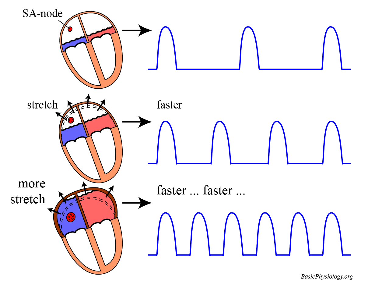
A. Intrinsic regulation: Sinus Node Stretch
1.
The sinus node is very sensitive to stretch. So, if the wall of the right atrium is stretched (by the presence of more blood), then the sinus node will also be stretched.
2.
When the sinus node is stretched, it will respond by initiating more action potentials.
3.
More action potentials will induce more excitations of the heart, and therefore more contractions.
4.
In other words, if the right atrium is more stretched because it is filled with more blood (from the veins), then the heart will beat faster.
5.
This is useful, as more blood will then be pumped out of the heart.
6.
This rapid pumping will, in turn, reduce atrial filling and the heart rate will then return to normal.
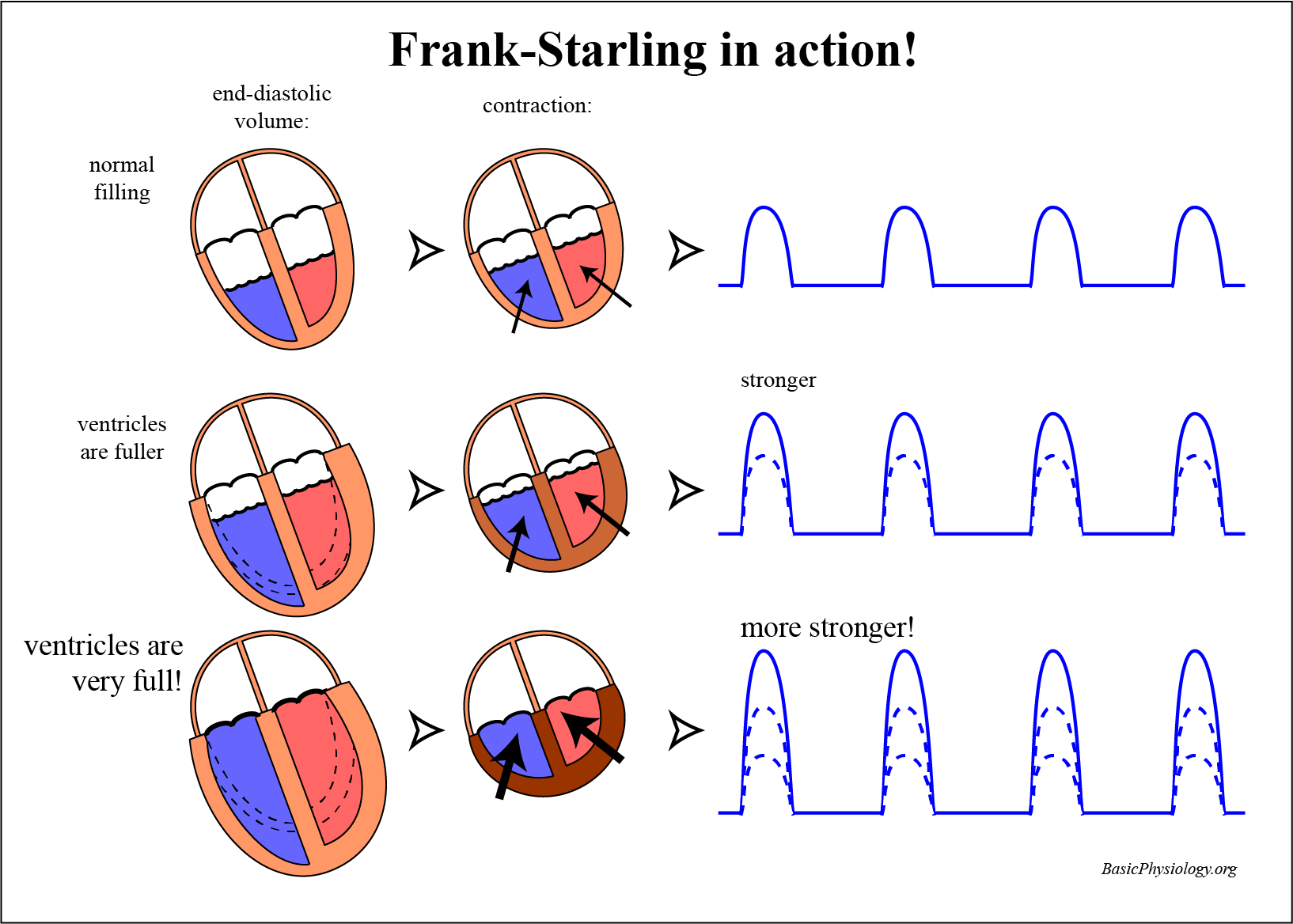
B. Intrinsic regulation: The Frank-Starling system
This mechanism is located in the heart muscle itself (intrinsic = belonging to the organ itself). It is a very important system!
2.
This system is named after the two people who first described this regulatory system (yes, you guessed it … Mr Frank and Mr Starling).
3.
Do you remember that, in skeletal muscles, when you stretched the muscle before the contraction, that this would increase the force of contraction? (link)
4.
The same mechanism applies to the heart. The more the cardiac wall is stretched, the stronger the next contraction will be.
5.
But, in the heart, the stretching of the cardiac wall is done by the blood that is accumulating in the chambers. The more blood in a chamber (atria or ventricle), the more the stretch.
6.
So, during diastole, blood flows into the ventricles, stretching the walls. The volume that is reached just before the contraction starts is called the end-diastolic volume.
7.
This system is very nice! If, for some reason, more blood flows into the heart, then the end-diastolic volume is increased, and this will (automatically!) induce a stronger contraction that will pump this extra amount of blood into the arteries.
8.
A stronger contraction means that more blood will be pumped into the arteries; i.e., the stroke volume will increase. Therefore, with this mechanism, the cardiac output (CO = SV*HF) has increased.
9.
The reverse is also true. If less blood flows into the heart, then stretch and contraction force will decrease and the cardiac output will be less.
10.
In summary, the heart, by itself (= intrinsic) adjusts itself to the amount of blood it receives to pump: if there is more, it will pump out more, if there is less, it will pump out less …
The sinus node stretch influences the frequency of the heart. The Frank-Starling system affects the contraction force.
2.
Since the frequency is the same for both hearts, the cardiac output of the right and the left heart are both influenced by stretching the sinus node (it is not possible for the right heart to pump at a different frequency than the left heart!).
3.
But in the Frank-Starling system, the end-diastolic volume stretches each ventricle and thus the contraction of the right ventricle could be higher or lower than that of the left ventricle.
4.
In conclusion, the Frank-Starling system, amongst others, keeps the balance between the right and the left heart.
As indicated in the diagram, there are two nervous systems innervating the heart; the parasympathetic and the sympathetic nervous system.
2.
The parasympathetic system (the vagus nerve) only innervates the sinus node and the AV-node. It inhibits the firing rate of the sinus node (longer P-P) and delays the propagation though the AV-node (longer P-Q).
3.
The sympathetic system also innervates both nodes, but also the myocardium itself, both in the atria and in the ventricles.
4.
An increase in sympathetic activity will:
Increase the heart rate
Increase the propagation velocity in the AV-node
Increase the contraction force in the cardiac muscle (atria and ventricles).
As an example of how the autonomic nervous system work on the heart, consider this very nice reflex: the Bainbridge reflex, also called the Respiratory Sinus Arrhythmia.
1.
The reflex is very simple: when the pressure in the right atrium increases, the heart beats faster. If the pressure decreases, the heart rate goes down.
2.
As the pressure increases in the right atrium, possibly due to an increase venous return, the atrial stretch receptors are activated which send their signals to the medullary centre in the brain. This in turn activates the sympathetic system.
3.
An increase in heart rate is then useful, as it makes the heart pump more blood, which will decrease the pressure in the atrium.
4.
You see this reflex very well during forceful in- and expiration.
5.
As you inhale, the pressure in the thorax decreases, which induces an increase in venous return and an increase in blood flow to the right atrium. This increases the atrial pressure -> increase heart rate.
6.
After inhalation follows expiration, the thoracic pressure increases, the venous return decreases, less blood flows into the atria, the pressure decreases and the heart rate decreases.