H.1. Introduction Nervous System

A. Introduction

1.

And now we arrive at the most central and complicated system in the body; the nervous system!

2.
This is the system that controls everything in our body; from the blood pressure, the digestion in our guts to our thoughts, our memories and, yes, our intelligence!

3.

In fact, this system is so complicated that ‘they’ have divided this ‘organ’ system into three parts:

  1. The Central nervous system
  2. The Peripheral nervous system
  3. The Autonomic nervous system

4.

The central nervous system consists of two major structures:

1. the brain

2. the spinal cord

These organs are so delicate that they have to be protected by the skull and the vertebral column.

5.
The peripheral nervous system is the nervous system that connects the central nervous system to all the organs in the body (heart, blood vessels, muscles, glands, etc). It is called ‘peripheral’ because this system is located outside the central nervous system.
6.
The nerves in the peripheral nervous systems can be divided into two systems:
  1. Afferent nerves: transport sensory signals from receptors in the body to the central nervous system.
  2. Efferent nerves: transport signals from the central nervous system to peripheral effectors such as muscles, glands etc.
7.
The autonomic nervous system is a system that controls all kind of functions in our body such as blood pressure, respiration, digestion.
8.
This autonomic system can (also!) be divided into two parts:
  1. The Sympathetic system
  2. The Parasympathetic system
9.
Although I will discuss this in more details later, one can say that the sympathetic system ‘stimulates’ local organs whereas the parasympathetic system has the opposite effect, makes them ‘quieter’.
B. Nerve cells

1.

What are the cells in the brain (and the spinal cord)? The most common cells are the nerve cells and the glia cells.


2.
We already discussed the nerve cells, in some detail, in a previous chapter (The Nerve Cell A.3.) but I will summarize the major points here.
3.
A typical nerve cell consists of the following four components:
  1. dendrites
  2. soma (= cell body)
  3. axon
  4. pre-synaptic terminals
4.
The dendrites are structures that ‘receive’ signals from other nerve cells.

The soma (=cell body) is where all the intracellular structures are located to keep the nerve cell alive (mitochondria, etc).

5.
The axon (sometimes short, but in some nerve cells very long; > 1 meter!) is an elongated tube that transports the action potential to the other end of the nerve cell.
6.

The pre-synaptic terminal is the ‘end’ of the nerve cell where the action potential can ‘jump’ to the next (nerve) cell.

C. The Action Potential
1.
The major function of a nerve cell is to produce and transport an action potential; which is an electrical signal. This is the signal that says “Hé, here I am!!”.
2.
This action potential can be generated by electrical signals originating from neighbouring cells, that are connected to this cell by a synapse.
3.

You may remember that at rest, a nerve cell displays a negative potential; typically, about -90 mV inside, compared to outside the cell (see A.3.2. The Resting Potential).

4.
When a nerve cell is stimulated, it produces an ‘action potential’. This is a sudden change in the potential, from -90mV to ±30 mV. This is called the ‘depolarization’.
5.

But once the cell is depolarized, it quickly restores its negative potential, back to about -90 mV. This is called the ‘repolarization’.

6.
In other words, the action potential is very short; only 5 to 10 milliseconds. A short but sharp signal!
7.
Some of you may remember here that action potentials can last much longer, especially in (cardiac) muscles; up to 300 milliseconds! (link).
8.
But what do we do with an action potential? Nothing, unless it is transported to another cell!
9.
Once an action potential is generated, it can either be transmitted to another cell (a chain reaction) or initiate something else such as a muscle contraction, gland secretion, etc.
D. Action Potential Transmission
1.
Transmission to another (nerve) cell can occur in two different ways:
  1. an electrical synapse
  2. a chemical synapse

2.

In this diagram of an electrical synapse, you can see that the two neighbouring cells are connected by small tubes, called ‘connexon’. These tubes allow the flow of intracellular ions such as potassium, to influence the potential in the neighbouring cells and initiate a new action potential.

3.

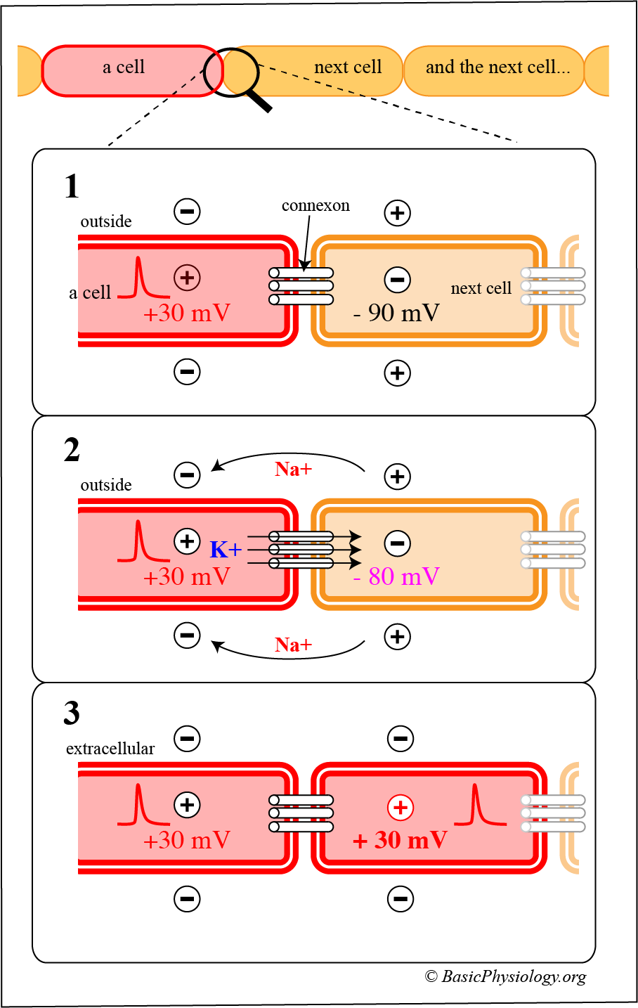
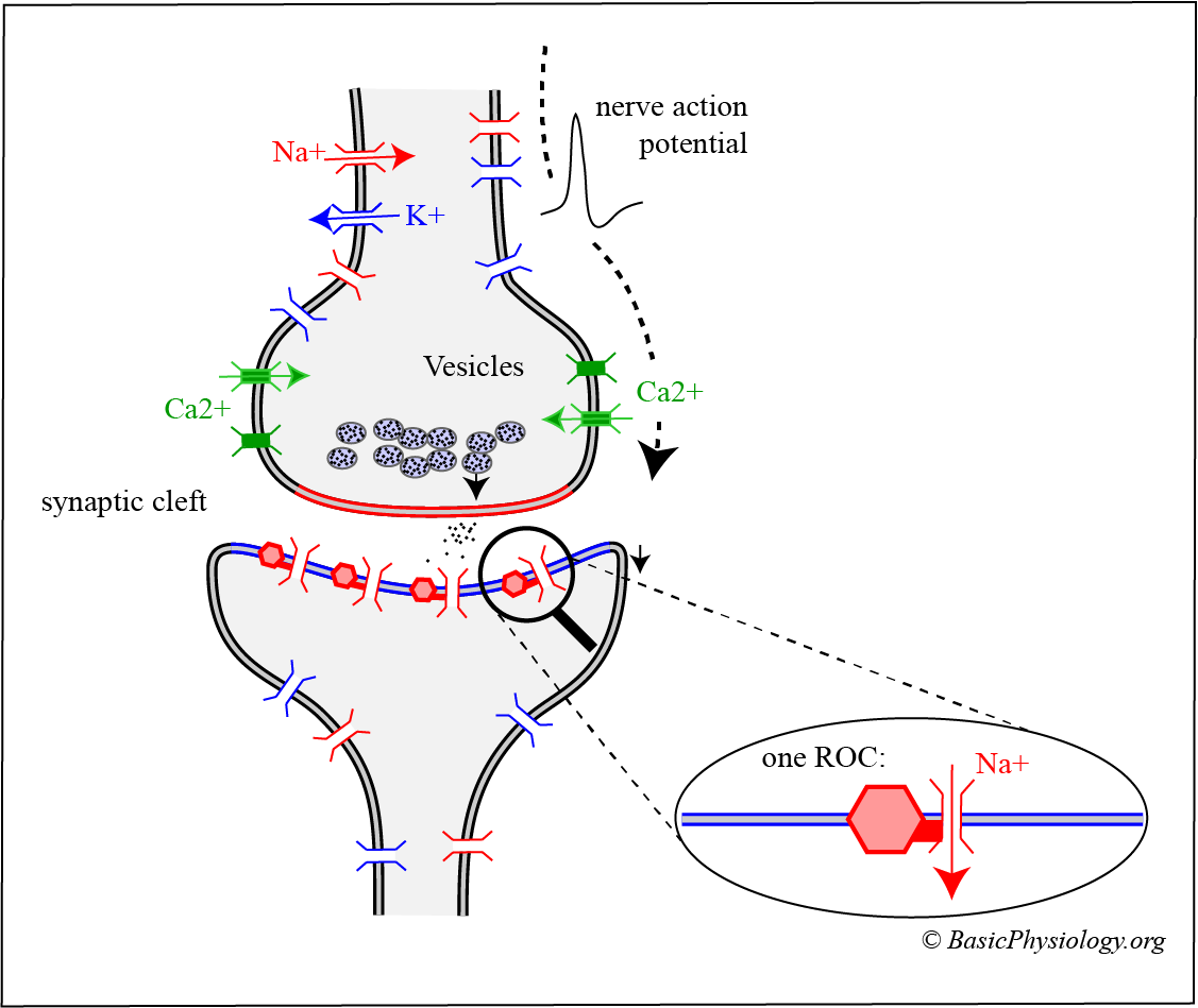
A chemical synapse is much more complicated, as you can see in this diagram. It consists of an interplay between the electrical signal, the function of a transmitter that has to diffuse through the synaptic cleft to the next cell, and the opening of new channels in that second cell. This all takes much more time than in the electrical synapse (about 1-4 milliseconds compared to 0,2 msec in an electrical synapse!).

4.
Furthermore, in contrast to the electrical synapse, the new signal is NOT an action potential but a small change in the post-synaptic potential.
5.

There are actually two different types of chemical synapses; an excitatory and an inhibitory chemical synapse, called an EPSP or IPSP.

6.

An EPSP (Excitatory Post Synaptic Potential) is a small depolarization in the past synaptic membrane. If several depolarizations are quickly initiated by incoming action potentials, then the threshold in the postsynaptic membrane can be reached and a new action potential is generated.

7.
In an IPSP (Inhibitory Post Synaptic Potential) synaps however, incoming action potentials will shift the post-synaptic potential further away from threshold making this nerve cell less excitatory. In a certain sense, these chemical synapses function like positive (or negative) calculators! We shall discuss later why such ‘calculations’ are crucial to our mind and body.
E. Glia cells
1.
Glia cells (also called neuroglia) are very different cells and their purpose is to support neighbouring nerve cells and their environment. They are (also) located in the central and peripheral nervous system.
2.

There are several types of glia cells with different functions, all of which will be discussed later (coming …!)

3.
Breaking news! Recently, a new system was discovered which describe and analyse the function of these glia cells; the Glymphatic System.

H.1. Introduction Nervous System

Slides