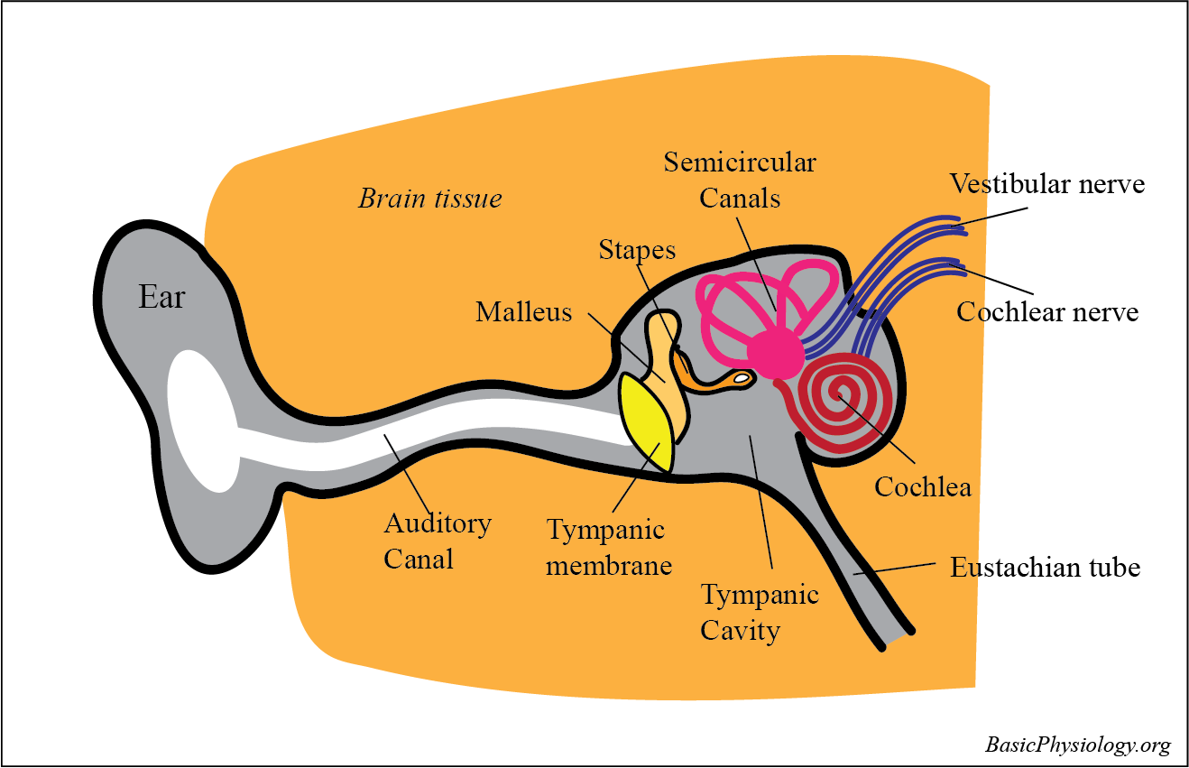
The ear is (like all special senses) quite complicated! It consists of three different parts:
the outer ear
the middle ear
the inner ear
2.
The outer ear is what we all can see; a beautiful shape that collects the sound signals from the outside world and which we sometime adorn with jewelry and/or piercings. And, it also contains the auditory canal that conduct the sound waves towards the tympanic membrane.
3.
The middle ear consists the three ossicles that collect the sound vibrations from the tympanic membrane, amplifies them and transmit to the inner ear
4.
The inner ear is actually the cochlea, a spiral shaped structure with a base, close to the middle ear, and an apex, at the distal end. This is where the sound signal is transformed into neural signals, to be transported towards the brain.
5.
What is confusing to many students is that adjacent to the cochlea, there is another structure, the semicircular canals.
6.
This is however a totally different system, part of the vestibular system, another special sensor, that detect and controls our body balance (see H.3.3.3. Balance).
In the air-filled space between outer ear and inner ear is the middle ear. Inside, there are three ossicles; the malleus, the incus and the stapes:
the malleus is connected to the Tympanic Membrane
the incus connects the malleus to the stapes
the stapes is connected the oval window.
These ossicles work aslevers; therebymagnifyingthe amplitude of the sound waves (approximately 20x).
2. The Eustachian Tube:
Connects the middle ear to the pharynx (inside your throat!) and therefore to the outside world. This communication is necessary to keep the pressure inside the middle ear equal to that outside the Tympanic membrane.
3.
Otherwise (if there is a pressure difference), this will cause the tympanic membrane to be pushed either in or out of the middle ear. This imbalance will make a persondeafand can be very painful.
4. The Attenuation Reflex:
The ossicles are very delicate and, when there is a loud noise, they need protection. Two muscles are attached to the ossicles:
1. thestapedius muscle
2.thetensor tympani.
5.
These muscles contract by reflex, when there is a loud noise, to pull at the ossicles and to prevent them of getting dislodged.
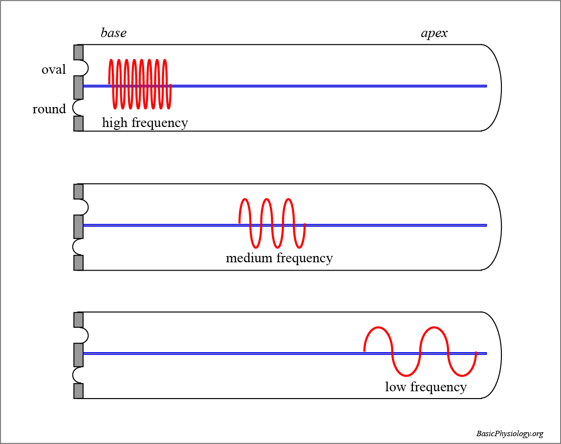
The cochlea is a spiral shaped structure with a base, close to the middle ear, and an apex, at the distal end.
2.
The inside of the cochlea is subdivided into three scala’s:
the scala vestibuli
the scala media
the scala tympani.
3.
The scala media and scala vestibuli work together. Between these two and the scalatympani, there is the basilar membrane.
4.
On this basilar membrane, the sensor of the ear, the organ of Corti is located.
(Scala = Italian for staircase)
5.
The inner earcan be described as a single tube which starts at the oval window and ends at the round window. This tube is filled with fluid. If the membrane of the oval window is pushed inwards, by a sound vibration, then the round window membrane, at the other end of the tube, is pushed outward. This is because fluid is not compressible.
6.
The opposite happens when the oval membrane is pulled outwards, then the round membrane will be pulled inwards. During normal hearing, this push and pull will occur very quickly and the fluid will oscillate left and right (thereby creating vibrations in the basilar membrane; not shown in this diagram).
7.
The basilar membrane will vibrate when the fluid in the upper and in the lower scala’s vibrate. But the basilar membrane is not the same (homogeneous) along its length. Rather, it contains fibers that are short and stiff in the base and long and slender in the apex.
Therefore, the basilar membrane will vibrate more at the base when the sound has a high frequency. Lower frequencies are better detected in the basilar membrane in the apex.
The usual way of hearing is by conduction of sound waves through the air to our ears. That is how we listen to music, to talks, to my lectures, etc.
2.
But, when we, ourselves,talk, then we also listen to ourselves in a different way, namely through bone conduction.
3. Bone Conduction:
As shown in the diagram, sound waves are generated in thelarynx and propagate from the mouth through the air to the ears. But they also propagate through the skull to the ears. This is called bone conduction. Therefore, we hear ourselves talking both through the air and the skull.
4. The propagation and the frequency of the sound through the skull is influenced by the bone. Therefore, we hear ourselves talk in a different way from anybody else. The only time you really hear your own voice is when you hear it back from a recording on your mobile, an iPad, or something like that. And then; nobody likes their own voice!