Page Links:
A. Introduction:
1.
As you can imagine, the large brains consist of several systems located in different parts of the large brains.
2.
We can not consider ALL systems (there are so many) but will concentrate on the most important ones:
- The motor system
- The sensory system
- The autonomic system????
3.
Of course, nothing is simple, certainly not in the brain. The motor system, for example, also consists of two different systems, fortunately collaborating with each other (!). These are:
- The pyramidal motor system
- The extrapyramidal motor system
1.
The function of the motor system, as you can imagine, is to determine our motility, for which we have the skeletal muscles, the muscles that are connected to our skeleton.
2.
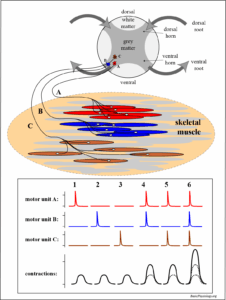
The skeletal muscle will contract when it is stimulated by an impulse (= an action potential) that comes from a nerve cell located in the spinal cord. This is what we called a motor unit (link: A.4.6. Motor Units).
3.
This is in contrast to other types of muscle cells such as the smooth muscle cell or the cardiac cell which are innervated (excited) in their own organs (see chapter A).
4.
5.
As you can see in this diagram, the action potential that will stimulate a group of cells in a skeletal muscle originate from nerve cells located in the ventral horn of the spinal cord; ‘a’, ‘b’ and/or ‘c’ nerve cell (and many more …)
6.
1.
As you can see in the diagram, the principle is quite simple; there is a nerve cell, located in the cortex (= superficial layer) of the large brain. The axon from this cell then conducts the nervous impulses all the way through the brain, through the medulla into the spinal cord until it reaches the appropriate level in the spinal cord.
2.
At this level, the axon synapses to a second nerve cell which is the cell that innervates the appropriate skeletal muscle fibber. That’s all!
Really?? Yes and no 😜!!!
3.
Yes; there is one specific neuron in the large brain cortex that sends signals, through a second nerve cell in the spinal cord, to stimulate a group of fibres in a specific skeletal muscle.
4.
And since we have a lot of skeletal muscles in our body, there must be a lot of nerve cells in the cortex that are all coupled to specific muscles.
5.
But the diagram also shows something even more strange. Look at the second axon (axon B). It also starts from the same area and runs through the large brain, but, just below the medulla, it suddenly ‘jumps’ to the other side and innervates a spinal cord cell in the right side of the body!
7.
This crossing-over occurs in a specific region of the medulla called the pyramids (one left and one right).
8.
That is why the axons that run through the pyramid and remain on the same side (±5%) are called the pyramidal tractwhile those crossing over (±95%) are called the extra-pyramidal tract.
1.
This diagram is essentially a repitition of the previous diagram but with large number of axons plotted to show the extend of the motor nerve cells in the cortex and their axons running through the brain and of course the large number (95%) of axons that cross-over to the other side. This crossing over is called ‘decussatio pyramidum’ which is latin for “crossing at the pyramid”.
2.
In the spinal cord, the two tracts are called tractus cortico-spinalis ventralis (same side) and tractus cortico-spinalis lateralis (opposite side). You don’t need a lot of latin to understand this!
3.
Ok, let’s go back to the cortex because there is something there which is even more interesting!
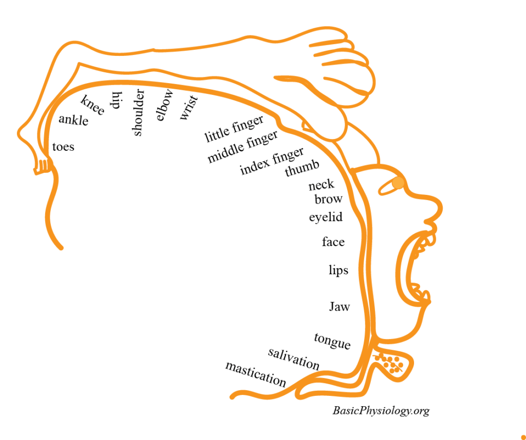
This diagram shows the relationship between the location of the motor neurons in the cortex and the body parts that are mobilized by these skeletal muscles.
4.
Now you can see which neurons innervate the muscles in the legs (toe, ankle, knee), innervate the muscles in the face (lips, jaw etc.), and more. As you can see, some areas, indicated by the black bars, are larger than others. For example, the number of neurons innervating the muscles in the lip is much higher than those innervating the muscles in the neck, for example.
5.
6.
Sometimes, this is called the cortical “homunculus”. This is Latin (again!) for a small human being, but distorted to reflect the degree of motor neuron innervation of the body parts.
7.
Another way to look at this cortex, is shown in this diagram.
Here you can see the location of the precentral gyrus with, in white, the location of the motor neurons innervating several major body parts, from the legs (top) all the way to the mouth (bottom).
8.
Btw, this is an important name to remember; precentral gyrus. Gyrus means a ridge-like elevation of the brain tissue. “Precentral” means “before the central sulcus”. The central sulcus is a groove that separates the precentral gyrus from the postcentral gyrus.
9.
Sorry for all these terms but these are quite common in this field; sorry!!


