What are reflexes? These are ‘automatic’ responses in the body. These can be motor or sensory responses, usually induced by a stimulus.
2.
For example, if you are in a dark room and see suddenly a bright light, then your pupils will react, automatically. Likewise, if your finger suddenly feels something sharp, the muscles in your arm will automatically jerk your hand away from this stimulus. This is called the withdrawal reflex.
3.
Reflexes are always very fast, and, you cannot control them.
4.
On this page, we will discuss the neuronal reflexes, that occur in the central nervous system. We will start with the most simplex reflex; the muscle stretch reflex.
B. Muscle Stretch Reflex:
1.
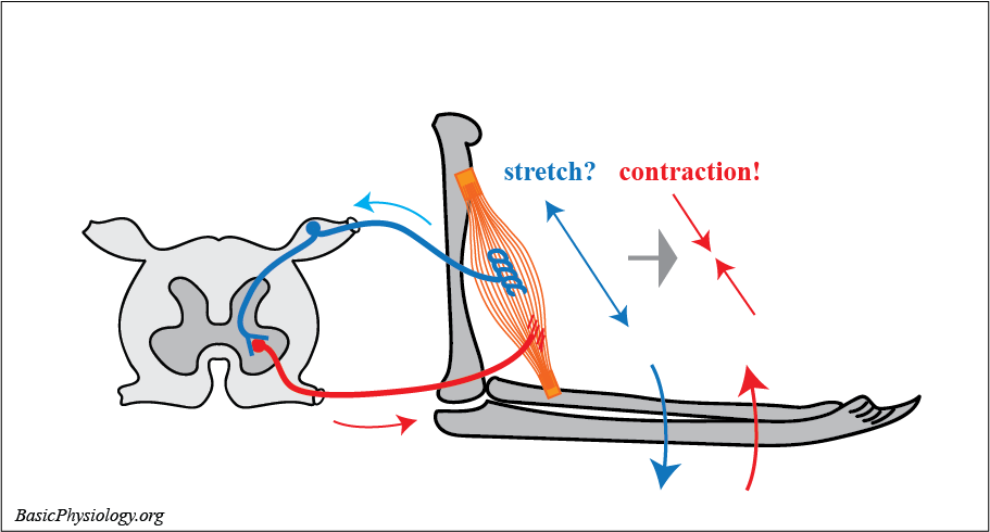
Probably the most ‘basic’ type of reflex is the stretch reflex, also called the myotatic reflex.
2.
As you can see in the diagram, it consists of a muscle spindle and a motor endplate, both located in the same muscle. From the muscle spindle, a sensory nerve propagates to the spinal cord where it enters into the dorsal root.
3.
There the nerve connects, in the anterior horn, with a synaps to an alpha-motor neuron. The axon of this neuron leaves the spinal cord, through the dorsal root, to the motor endplate of the same muscle.
4.
Its function is simple. When the muscle is (suddenly) stretched, this stretch is detected by the intrafusal fibers in the muscle spindles.
5.
This stretch induces action potentials in the muscle spindle that propagate, through the sensory nerve to the spinal cord. The nerve ends as a synapse onto the body of the motor neuron, in the anterior horn.
6.
There it stimulates the neuron to produce action potentials that propagate, through the motor nerve, to the motor-endplates of the same muscle.
7.
This will lead to muscle contractions that will reduce the length of the muscle to its original length. It is like a feed-back loop!
8.
This reflex works very fast! From stretch to contraction, about 25 msec!
C. Body Position and Posture:
Why do we need this stretch reflex?
2.
To keep our posture! Without this reflex, our body would collapse.
3.
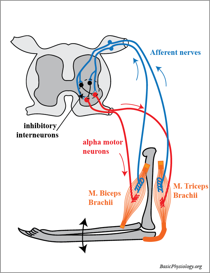
Look at this simple diagram. The biceps muscle holds the lower arm at an angle to the upper arm, in this case about 90 degrees.
4.
If for some reason, the lower arm moves downwards, then the intrafusal fibers in the muscle spindle will also be stretched which will evoke action potentials.
5.
These action potentials will propagate to the spinal cord where it will excite the alpha-motor neurons of the biceps.
6.
These action potentials will propagate back to the biceps to increase its contraction force which will move the lower arm back to its original position. All this automatically and very fast (25 msec!).
7.
But what about the other muscles that are also attached to the arms around the elbow joint? After all, they also play an important role in keeping up this position.
8.
But, as you can see in this second diagram, it now becomes a bit more complicated in the spinal cord.
9.
As one muscle, for example the biceps, is contracting, then the ‘opposite’ muscle, the triceps, must do the opposite; relax!
10.
And if the triceps contracts, then the biceps must relax. And all this very very fast!
11.
Fortunately, this is taken care of by negative intermediary neurons (in black in the diagram). So, when one alpha motor neuron is activated, the ‘opposite’ motor neuron is inhibited.
12.
And, as you have to realize, this is the case for all our skeletal muscles in our body, adjacent to all our joints, from elbow to knee joint etc.
D. Knee jerk reflex:
1.
A nice way to test this system, which is often used by doctors, is the knee jerk reflex.
2.
In this test, a hammer is used to strike the tendon of the knee patella.
3.
This tendon is attached to the upper leg muscle. The sudden ‘jerk’ of the tendon will suddenly stretch the muscle fibers including the muscle spindles inside that muscle.
4.
As the intrafusal fibers are also suddenly stretched, they will send action potentials along their afferent nerves to the spinal cord.
5.
This will excite the corresponding alpha motor neuron which will contract this muscle and move the lower leg, suddenly, upwards, like a ‘kick’.
6.
At the same time however, the opposite muscle must be inhibited (relaxed) to allow this jerk movement to take place.
7.
Therefore, that muscle must be inhibited. This is performed by an interneuron, located in the spinal cord and activated by action potentials from the same excited muscle spindle.
8.
However, the action potentials in that interneuron will inhibit action potentials of the opposite muscles, allowing that muscle to relax.
E. Withdrawal reflex:
1.
The withdrawal reflex occurs when your hand (or foot) encounters a sharp (or hot) object inducing a sudden withdrawal of your limb.
2.
The purpose of this reflex is to jerk your hand away from this dangerous stimulus.
3.
And again, this must happen very fast, to avoid further damage to your hand.
4.
The diagram shows the most important pathways of this withdrawal reflex. Starting with the hand where a sharp object has stimulated a painful stimulus.
5.
This induces action potentials in the sensory nerves that travel to the spinal cord. Once in the spinal cord, the nerve synapses to a motor neuron and to an interneuron.
6.
The motor neuron is connected and excites the corresponding flexor (in this case the biceps) to contract strongly.
7.
The interneuron is connected, as an inhibitory neuron, to the motor neuron of the opposite muscle; the extensor.
8.
This is important because you want the arm to move rapidly away from the painful stimulus and therefore, the extensor muscle must not contract, but relax.
F. Cross-over reflex:
1.
In the arms, the withdrawal reflex works fine; jerk your hand and arm away from the painful stimulus. That’s all!
2.
But in the legs, jerking your foot suddenly away from a painful stimulus is more complicated.
3.
If your right foot/leg is suddenly jerked away, then you would suddenly fall over! And, remember, all this happens very quickly.
4.
To avoid your falling over, your other leg must take over your posture. Fortunately
5.
But since several muscles in the upper and lower legs are involved in this reflex, the neuronal connections must also cross-over to the other side of the spinal cord and to other segments in the spinal cord.
6.
So, the flexor in the right leg must be stimulated to withdraw the foot from the painful stimuli. This is a simple withdrawal reflex.
7.
At the same time, the opposite extensor muscle in the right leg must be inhibited. Like in the withdrawal reflex
8.
But, the muscles in the left leg must also be excited. But this time it is the extensor that must be stimulated while the flexor must be inhibited. The opposite action!
9.
Fortunately, as you can see in the diagram, there are several interneurons that connect the motor neurons in the right way.
10.
Btw, the withdrawal effect is only functioning in the legs. Not in the arms! We don’t need that in the upper limbs.
11.
That’s because you will not ‘fall over’ when you suddenly ‘withdraw’ an arm. The legs are keeping us stable, thanks to all the stretch reflexes in our legs.